December 18, 2024
Building a new surgery center requires a design team to ask one key question every step of the way: “What will this look through the eyes of the patient?”
This website uses cookies. to enhance your browsing experience, serve personalized ads or content, and analyze our traffic. By clicking “Accept & Close”, you consent to our use of cookies. Read our Privacy Policy to learn more.
By: Danielle Bouchat-Friedman | Senior Editor
Published: 10/3/2022
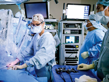
Facilities with the foresight to invest in the blending of surgeon skill and technological advancements are shaping the future of surgery. MultiCare Health System in Tacoma, Wash., added the da Vinci Xi surgical robot system to three of its 13 ORs. The robots are more than flashy marketable technology to Laila Rashidi, MD, FACS, FASCRS, a colon and rectal surgeon at MultiCare Health. She says the enhanced 3D vision and advanced instrumentation capabilities have helped her perform less invasive and more precise surgeries. “Having access to the surgical robot has changed the way I operate,” says Dr. Rashidi, recalling how much colon and rectal surgery has changed since her fellowship training in 2016. “Colectomies have long been inpatient procedures, with a length of stay averaging three to five days. Now, I safely perform the procedures on appropriately selected patients in the same-day setting.”
Robotic assistance allows Dr. Rashidi to perform less invasive surgery than what’s possible with the laparoscopic approach, resulting in significant reductions in operative times, recoveries and cost of care. “With the right preoperative planning,” she says, “most of my colectomy patients go home the day of surgery, or the next day, rather than having to recover in the hospital.”
Operating laparoscopically can be limiting, requiring more manipulation and ultimately more tissue trauma, points out Dr. Rashidi. “With the robot, I can use three arms that have a greater range of motion and dexterity than my own hands,” she says. “I feel completely in control when I’m operating, and when you’re fully in control your surgery will go faster and results will be better.”
Before procedures, Dr. Rashidi logs into her account in the robotic platform and engages her preferred ergonomic settings for the surgeon’s console. “It’s a huge relief to sit comfortably when you’re operating for as many as seven hours at a time,” she says. “The neck cramps I used to suffer while operating laparoscopically have nearly vanished.”
The automated set-up saves time before each case because Dr. Rashidi can access her preset positions based on the procedure she’s about to perform and her preferred approach. Each surgeon who performs robotic procedures at MultiCare Health System has their own account and can set their preferences based on specific procedures.
During surgery, Dr. Rashidi views high-definition and magnified 3D images of anatomy and tissue. The robot’s articulating arms bend and rotate to move instruments with incredible precision, which allows for better tissue handling, less bleeding, limited trauma to internal structures and less postoperative pain.
The robot is equipped with advanced visualization technology such as 3D modeling, which lets Dr. Rashidi preplan procedures based on CT scans of the patient’s anatomy. She can review 3D models of the anatomy before surgery, input the surgical plan into the robotic platform and refer to it throughout the case. The robot’s integrated fluorescence imaging capability employs near-infrared technology, which allows Dr. Rashidi to identify critical structures and evaluate bowel perfusion prior to anastomosis.

Dr. Rashidi and her colleagues are waiting on a digital component upgrade to be installed that will give them the ability to store and share video and images from their procedures. “I’ll be able to easily access video recordings of my surgeries on my phone or computer and review each step,” she says. “I like to look back at each of my cases to see if I made extra or unnecessary moves with the robot. I’m a big believer in watching videos of my procedures because I learn so much and can improve my approaches for future operations. Reviewing previous successful cases also increases my chances of reproducing the same positive outcomes for patients.”
Dr. Rashidi edits her own videos for teaching purposes and the new software system will help her to easily find the sections she wants to highlight. “I’ll also be able to share my videos with other surgeons,” she says. “I find it helpful to get another professional perspective to learn and improve.”
Through an app on her phone that is synchronized to the robotic console, Dr. Rashidi can access and study data from procedures she previously performed and can even compare her performance to national benchmarks. “I love being able to access
the app on the robot’s console or my phone because I can easily pull up relevant information,” she says. “The technology enables me to constantly learn, improve and analyze critical data while operating with more precision
and efficiency.”
Many years ago, the thought of inserting a tiny camera through a small incision to perform surgery may have sounded far-fetched, but today minimally invasive arthroscopic surgery is one of the most
commonly performed procedures in the U.S. Now, orthopedic surgeons can look forward to another innovation that will improve how they scope joints: a wireless camera system.
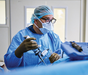
Laith M. Jazrawi, MD, performed a knee arthroscopy last month at NYU Langone Orthopedic Center in New York City using a wireless camera that recently received market clearance from the FDA. The wireless camera runs off a battery, which, when fully charged, can last one hour and 15 minutes. “For routine procedures, that’s plenty of battery life, but it can always be changed out quickly if necessary,” says Dr. Jazrawi.
He points out that surgeons often fear that wireless video technology could cause lag time in the direct transfer of the image. “There’s no lag with this system, so whatever I am looking at with the camera shows up on the monitor in real time,” says Dr. Jazrawi.
Wires and cords present certain limitations in the OR by tethering surgeons to equipment and could also cause sterility concerns, according to Dr. Jazrawi. “The less that you are connected by cables, the faster you can turn over rooms and the quicker you can start the next case,” he says, pointing out that the wireless system also allows for more ergonomic and efficient surgical movements.
Dr. Jazrawi says the truly exciting thing about the wireless camera system is that it creates the possibility of untethering other surgical equipment. “Perhaps we’ll be able to cut the cord for the shaver next,” he says.
When Essentia Health in Duluth, Minn., broke ground on the Miller Hill Surgery Center, including integrated surgical suites was part of the plan. The center, which opened in August, features four 600-square-foot ORs equipped with
state-of-the-art technology, including an integration system that is easily accessed with touch panel controls.
Each OR is equipped with 55-inch video monitors that display crystal clear surgical images and X-rays of the patient’s anatomy for the entire surgical team to view. The images are easily uploaded to the electronic health record, which providers can access during future consults with patients. A surgeon’s preference card can also be pulled into the integration platform and displayed on the monitors so the surgical technologists know exactly how the room should be set up and how the instruments on the back table should be arranged.

Although OR integration technology is a draw for staff and surgeons, Stacy Lund, operations director for acute surgery, says its purpose and priority is to maintain patient safety. “The new surgery center is not connected to our main campus, so we wanted physicians in the space to have the ability to contact other providers throughout the health system during procedures,” says Ms. Lund. “The integration platform allows us to video conference with physicians and engage in peer-to-peer consults in real time.”
The ORs have a zero-footprint design, which means video equipment is located outside the room and accessed through the integration platform. This provides a safer, more sterile operative environment, according to Jefferson Davis, DO, an orthopedic surgeon at Essentia Health. “The integration clears up a lot of the cords and equipment that tends to clutter the OR, making it much easier to navigate around the patient without worrying about slips and trips and bumping into things that aren’t sterile,” he says.
Despite being cutting-edge, the integration technology is very user-friendly. “The beauty of this system is that it is fairly seamless and doesn’t require specialized training to operate,” says Dr. Davis.
The recently renovated Eisenhower Desert Orthopedic Center (EDOC) in Rancho Mirage, Calif., now boasts eight 595-square-foot ORs filled with robotic platforms, two 80-inch wall-mounted ultra-high-def video (UHD) monitors and three 36-inch UHD monitors positioned
on booms around the surgical table. “We’re monitor-heavy,” laughs Stephen J. O’Connell, MD, a fellowship-trained and board-certified orthopedic surgeon specializing in surgery of the hand, wrist and shoulder at EDOC.
The numerous screens positioned strategically around the room let Dr. O’Connell and his colleagues refer to preoperative and intraoperative images in ergonomically and efficient ways at any point during procedures.
He recalls craning his neck to see arthroscopy video displayed on a monitor less than ideally positioned around the sterile field or trying to decipher anatomical images on a small screen attached to a C-arm while trying to drill in a pin with pinpoint accuracy. Now those images are seamlessly routed to the large monitors in the room. “No matter where I’m working on the patient, I can look up directly at a screen,” says Dr. O’Connell.
The integrated system connects to the facility’s EMR and fluoroscopy system, so any video or image captured during the patient’s episode of care — the renovated EDOC also houses an on-site clinic, radiology suites and physical therapy office — is automatically loaded into the facility’s PACS system or patients’ medical records for comprehensive storage and easy access.
After procedures, Dr. O’Connell steps into a private room adjacent to the ORs and uses an iPad to immediately access the case’s information, including videos and images captured through the integrated system. He can edit and annotate the videos and images and record a short video message to patients. During the short videos, he reviews the steps he took during surgery and includes images to support his explanations. He can also go over specific post-op instructions for patients to follow after discharge and remind them to schedule their two-day follow-up appointment.
Dr. O’Connell then emails the multimedia package to patients, who can review it when they get home and refer to the information as needed. Patients can also send him messages through the EMR’s portal if they have questions about the progress of their recoveries. “It’s a fully integrated system for medical records and imaging that can be easily shared with patients and healthcare professionals,” says Dr. O’Connell.
He’s proud of how hard he and his partners worked to turn the concept of a facility renovation into a state-of-art reality, and is excited to know the center’s wired ORs will keep pace with what’s possible in same-day surgical care. “It’s been an honor to be part of the project and it’s gratifying to know we’re set up for many years of future surgical success,” says Dr. O’Connell. OSM
Building a new surgery center requires a design team to ask one key question every step of the way: “What will this look through the eyes of the patient?”
Anthem Blue Cross and Blue Shield has reversed course on its plan to change the way it reimburses for anesthesia care payments, which critics said included not paying for...
It’s a crazy time of the year as the holidays are in full swing. Your team members may be extra stressed with their workloads and personal obligations, and you’re in the same...