.svg?sfvrsn=be606e78_3)
- Home
- Article
The Latest in Ophthalmic Surgical Microscopes
By: Joe Paone | Senior Editor
Published: 8/8/2024
Share:
A wealth of digital features provide enhanced clinical capabilities, increased collaboration and vastly improved ergonomics.
The surgical microscope has long been a crucial procedural tool in ophthalmology. Now, in a world where almost every object has a digital component that opens up a wide array of new capabilities to the end user, surgical microscopes for eye procedures are no exception.
Microscopes that leverage digital technology provide benefits that are undeniable. And for facilities whose budgets may be too tight to purchase new digital-enabled microscopes, the good news is that it’s possible to add some digital functionality to many conventional analog microscopes.
So what exactly are these benefits? Here are the primary ones.
Heads-up displays
Surgeons who use conventional microscopes spend days, weeks, months and years in uncomfortable positions, hunched over the eyepiece lens to perform incredibly precise operations. It’s a widely acknowledged fact that many ophthalmologists work with pain in their necks, backs and shoulders — a circumstance that has shortened many vibrant careers. We took a deep dive into the brutal ergonomics situation for eye surgeons last year.
“You’re never really in a comfortable position no matter how hard you try, and it can lead to chronic musculoskeletal issues,” says Robert J. Weinstock, MD, a board-certified ophthalmologist and fellowship trained cataract and refractive surgeon who serves as director of cataract and refractive surgery at The Eye Institute of West Florida and Weinstock Laser Eye Center in Largo. “Between the height of the bed, the height of the chair, the tilt of the microscope and the oculars and the pupillary distance you need to set, it’s never a super-comfortable position ergonomically when you sit down and operate. It’s a distraction if you have any pain, which can lead to you not seeing as well and not concentrating as much as you should be on the eye in the surgical field.” Another drawback of traditional microscopes, he notes, is the eye strain surgeons can suffer from looking at bright light through the eyepiece, especially at high magnification.
Dr. Weinstock, who also serves as medical director of the Largo Ambulatory Surgery Center, has long embraced the cutting edge of innovation in the realm of surgical microscopes and, given those significant drawbacks of conventional microscopes, he finds the heads-up display one of the most welcome of such enhancements. “The value of a tool that lets you operate in a more ergonomic and safe position is pretty valuable and pretty worth it,” he says. “When you’re back away from the microscope looking at a screen four feet away, you can mag up much higher without feeling that strain and headache.”
Dr. Weinstock says the screens, preferably in the 50-inch range, can be mounted on a boom or cart, with a station below that houses the computer that processes and augments images and can even record procedures. “You can twist the screen, move it and angle it, and roll it around the OR,” he says.
Increased OR team collaboration
By moving visualization from the eyepiece to a large screen, a digital-enabled microscope doesn’t just alleviate ergonomic issues. It also allows the rest of the surgical team to see exactly what the surgeon sees and follow what they are doing in real time.
“The conventional microscope is a one-man machine,” says Dr. Weinstock, noting that the heads-up display enables the entire OR to work more efficiently and proactively. “Because the scrub tech and the circulator can see the screen too, they know where we are in the case. They know when we’re about finished, when we’re putting the lens in, when we are having trouble because they can see it and anticipate what’s happening. There’s a collaborative effect that is subtle but adds to efficiency and, ultimately, better patient care.” Ultimately, with a heads-up display, staff is better prepared for what the surgeon is going to need in certain situations, which allows them to stay ahead of the curve.
“They can get things on the field faster to help the surgeon so he manages complications appropriately and quickly and can ‘save the day,’ so to speak,” says Dr. Weinstock. “They see the surgeon starting to struggle, going slower. They see the pupil is small. They get a heads up that something’s going on, and start in their own mind doing a checklist, getting the stuff they need, looking in cabinets, getting things ready just in case they need it. For example, if a surgeon is having trouble sealing the wound, they can see that on the screen and get the suture set ready over by the scrub tech so they’re ready to open it.”
It’s possible to add heads-up display capability to conventional microscopes. That’s the case at Eye 35 ASC in Schertz, Texas. “We use the conventional scopes, but have added a camera to record cases and to broadcast to the wall-mounted TV in the OR so everyone can see what the surgeon is seeing,” says Clinical Director Carson McCafferty, MSN, RN, CNOR, CNAMB, CSRN. “That helps the staff know what part of the surgery he is on so we can be efficient with our flow.”
For Eye 35, adding heads-up display capability to existing microscopes is a winning formula for the time being. “Our surgeons love the microscopes we have, even though they are a little old,” she says. “I think they are trying to get all they can out of them since they are happy with them. There is newer technology out there they probably will invest in before we invest in new microscopes.”
3D, augmented reality, real-time data
This is where digitalization really shines by providing a wealth of improved visualization capabilities to the surgeon, including enhanced detail, more convenient control of video settings, real-time on-screen data and information, and digitally augmented 3D views that highlight relevant structures and blood flow.
“Depending on what you’re doing, they have different filters you can apply that highlight different tissues,” says Dr. Weinstock. “If you’re doing a MIGS case, there’s a setting for that. If there’s blood in the eye you’re trying to remove, there’s a setting for that. If you’re trying to do the capsulotomy for a red reflex, there’s an optimization setting for that. Because they can use digital filters to enhance the image and you can turn up the contrast or gain, it also allows you to operate under lower light, so the patient’s not getting exposed to so much microscope light. That’s a little safer on the patient, and it allows for a little faster visual recovery.”
Another aspect of digital visualization that Dr. Weinstock lauds is picture-in-picture, particularly the ability to import diagnostics and other devices into the viewable surgical field. “If you have intraoperative aberrometry, for example, or preoperative information you want to check out during surgery, you can have a different source import information into the OR that you can see in the corner of the screen,” he says. “Some systems have guidance for aligning a toric IOL [intraocular lens] with digital graphics that are superimposed on the screen with tracking. Intraoperative OCT [optical coherence tomography], used by retina specialists, is pretty interesting. They can see a live OCT while they’re operating to help guide their surgery.”
Everyday benefits
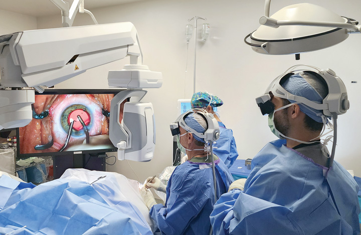
COLLABORATIVE EFFORT Dr. Robert J. Weinstock (seated) combines use of an augmented reality headset with heads-up visualization for the rest of the OR team, including recently graduated fellow Dr. Caleb Shumway (right). | Renee Bondurant
Victoria Wiltshire, MBA, RN, is vice president and chief operating officer of SightTrust Eye Institute, which runs a cataract and refractive lens replacement surgery center in Sunrise, Fla. She says digital microscopy has “revolutionized our medical practice” clinically, financially and operationally. The digital suite of equipment and applications the center uses enables surgeons to perform cataract and refractive lens replacement surgeries with improved precision, optimal outcomes, minimized complications and enhanced patient satisfaction that leads to positive word-of-mouth referrals, increased patient volume and, ultimately, improved revenue generation, says Ms. Wiltshire. SightTrust’s digital microscope system integrates with a biometer device and digital marker laser, which she calls “a game changer for our practice that streamlines surgical workflow and increases accuracy. These devices provide real-time, accurate measurements such as corneal thickness, anterior chamber depth and axial length, which are essential for precise IOL selection and placement.”
Ms. Wiltshire says SightTrust’s digitally enabled microscopes enable more efficient and accurate IOL selection by more precisely measuring each patient’s unique ocular anatomy. “We can confidently select the most suitable IOL, minimizing the need for costly IOL exchanges,” she says. “This optimization of the surgical process reduces overall costs and improves resource allocation within our center.” SightTrust’s system also enhances operational efficiency by seamlessly transferring surgical plans to the OR.
SightTrust additionally benefits from digital microscopy through the consistent flow of pertinent data into and out of the OR. “The integration of these technologies eliminates the need for manual data entry and minimizes the potential for human error,” says Ms. Wiltshire. “Measurement data is automatically transferred to our electronic medical record, ensuring accurate and up-to-date patient information. This streamlined approach saves valuable time for both our surgical team and administrative staff, allowing them to focus on providing high-quality patient care and improving overall productivity. The integration of these advanced technologies has facilitated effective collaboration and communication between our surgical team members. Real-time sharing of measurement data and imaging allows for seamless coordination during surgical procedures, leading to improved teamwork and enhanced surgical outcomes.”
What’s to come
Digital innovation in ophthalmic surgical microscopy continues. For example, Dr. Weinstock’s facility recently brought in a system that provides him a magnified 3D image of the surgical field with integrated preoperative information through an augmented reality headset. “It’s very immersive,” he says.
The headset provides head gesture control as well. For example, if he moves his head up or down, the system can automatically raise or lower the light, or control zoom or focus. “It allows you to use your head motions, which are much more delicate and sensitive than using your foot with traditional microscope pedals,” he says. “You can refine your image a lot quicker.”
Dr. Weinstock says the industry is just scratching the surface of what’s likely to be possible in coming years. He foresees unprecedented collaboration where surgeons wearing headsets can collaborate on surgeries from anywhere in the world. “There’s a huge ability to improve patient care with that type of collaboration, without the surgeon needing to actually be there,” he says, adding that this technology will provide benefits for educating surgeons on new techniques.
In his view, the sky is the limit once the microscope platform becomes digitized. “Anything that’s digital-related could be integrated,” he says.
Where we’re at
For now, eye surgeons are still absorbing and adapting to this early wave of digital microscopy. “It’s still the first inning. It’s way early,” says Dr. Weinstock. “Only the early adopters and innovators are working with digital microscopes and visualization, but it’s starting to become more mainstream. People are loving it, and more and more younger surgeons want it.”
Dr. Weinstock urges more experienced surgeons to take a fresh look at the new wave of products and capabilities. “The barriers of optics, latency, image quality, the functionality and robustness of the unit, the footprint — all these things people complained about in the past — I think those have all melted away,” he says. “It’s really a lot more plug-and-play than it used to be. Now it’s just a matter of finances.”
Digital microscopy is not for everyone — yet. Many veteran ophthalmologists are happy with their traditional microscopes, and some may be averse to new technology. Tech-savvy surgeons of all experience levels, however, will likely see the appeal. “If you’re a surgeon who has advanced their career and has implemented new technology into the OR and into your life, you’re not going to have a problem,” says Dr. Weinstock. “You’ve been through this before.”
As far as cost being a prohibitive factor, Dr. Weinstock says these upgrades might not be as pricey as leaders might think. “There are different ways you can do it,” he says. “You can add on to an existing microscope and just get the screen and the camera,” he says of one system. “You don’t need to buy the whole new base of the microscope and optical head.” In addition, some systems are modular, allowing centers to only pay for functionality they need, be it intraoperative aberrometry or OCT, or built-in guidance. “If you’re building a new ASC and buying new microscopes anyway, it’s probably not that much more cost than buying a conventional microscope,” he says.
For Dr. Weinstock, perhaps the most important feature of digital microscopy that he stresses to his colleagues is ergonomics. “It’s an absolutely critical investment in your career and in your health, and it’s very hard to put a price tag on that,” he says. “Do you want a long career? It just seems like a really smart investment in your health and your future long-term, and would reduce the chance of you having a career-threatening scenario come up when you are in your 50s or 60s from operating through a conventional microscope for that long a period of time, with a frozen neck or shoulder, chronic low back pain or disc issues. You can’t underestimate the value and the potential impact something like this could have on your career and your financial livelihood.” OSM
.svg?sfvrsn=56b2f850_5)