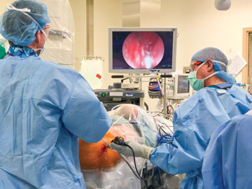
Surgeons at Stanford Health Care are performing the osteochondral autologous transfer system (OATS) procedure, which involves taking cartilage plugs from one part of the knee and using them to repair damaged cartilage on the other side of the
joint. Cartilage is limited in the knee, so the plugs must be 10mm wide or less, notes a report in Stanford Health Care Now.
Marc Safran, MD, chief of sports medicine at Stanford (Calif.) Health Care, addresses this limitation of the OATS procedure by transplanting patellar cartilage from a cadaver, an approach that fills the defective area of the knee with a single,
smooth piece of donor cartilage that can be sized appropriately. “What’s going to be key with the older population is to prevent the diseases of aging,” Dr. Safran told Stanford Health Care Now. “To me, that’s
the Holy Grail. Everyone is looking to solve or prevent arthritis.”
Dr. Safran and his colleagues are studying the effect excessive motion of the femoral head within the acetabulum has on hip microinstability with the aim of improving outcomes of hip arthroscopy, according to Stanford Health Care Now. The
outlet says he’s also championing the creation of the North American hip arthroscopy registry, which would collect patient data such as BMI and procedure type with the aim of analyzing hip arthroscopy outcomes and learning more about
the limitations of the procedures. His goal is to collect data from about 8,000 patients each year and capture reliable data over the next few years.
The benefits of emerging arthroscopic procedures are clear, but no change happens without some hurdles along the way. For surgeons, there can be a steep learning curve involved in adopting arthroscopic techniques and technologies. “The biggest
challenge for a lot of newer surgeons is getting the experience and the repetitions they need, and performing arthroscopic procedures until they become second nature,” says Dr. Bjerke. “Different types of cases can be challenging
from a technical standpoint.”
Dr. Getelman agrees that arthroscopic procedures are technically demanding. “Arthroscopy is a learned skill that takes years of practice to perfect,” he says. He adds that efforts to achieve arthroscopy success begin when the patient
is first evaluated in the clinic — a comprehensive patient history, detailed physical examination and clear imaging allow surgeons to make the correct diagnosis and address the underlying pathology of the patient’s condition.
Different types of cases can be challenging from a technical standpoint.
— Dr. Brian Bjerke
“Understanding the subtleties is the foundation to achieving successful outcomes,” said Dr. Getelman. “Surgeons must then anticipate and plan the surgical steps and have all necessary equipment available. The superior surgeon
will be methodical and efficient in their approach using reproducible and deliberate steps to treat each source of pathology.”
Dr. Urse believes quality pre-op imaging shows clear views of both femoral heads in the hip and provides a good view of the alignment of the sacrum and the top of the pubic symphysis. “Surgeons can notice abnormal or well-formed sockets
to determine if hips are dysplastic,” he says. “It allows us to find out why the patient is experiencing joint pain.”
In the OR, applying the proper amount of traction to the patient’s leg during hip arthroplasty is key to achieving excellent outcomes. Patients are most often placed in the supine or lateral position, says Dr. Urse. Most surgeons perform
the procedure with the patient in the supine position with traction applying pressure to the upper medial thigh to get lateral displacement. Slight abduction and a bit of flexion relaxes the capsules, according to Dr. Urse. “Traction
is the key to surgery. It lets you open the joint to get the views you need to perform the procedure,” he explains. “As Dr. Jim Glick, an early pioneer of arthroscopic techniques said, ‘Inadequate traction is the root of
all evil in hip arthroscopy.’”
Dr. Getelman says surgeons still need to be prepared to convert arthroscopic procedures to open surgery when the minimally invasive approach isn’t feasible for medical or technical reasons and cautions against surgeons becoming too reliant
one way of performing surgery. “Arthroscopic techniques can be a great positive, but it can be a negative if that’s the only way you know how to repair a joint,” he says. “Surgeons must learn the basics and the fundamentals
of the surgery in order to take full advantage of technology as a tool to improve it.”
Dr. Bjerke believes high-tech arthroscopy procedures should be seen as a complement to a surgeon’s existing skillset — a way to perform complex techniques in a safer or less invasive way. He adds that it’s easy for surgeons,
especially younger ones who have always had these options, to get too reliant on the technology.
“Surgeons do things they’re comfortable with and they get into a routine, which is good. You want to be able to do things well and repeat them,” he says. “But not being able to perform a specific surgery because a certain
supply or instrument isn’t available is a red flag, indicating surgeons need to expand their repertoire and knowledge of what else is available.”
That education needs to be included for other surgical staff, too. In many cases, if something goes wrong, it’s the nursing staff or surgical techs who must troubleshoot the equipment. The surgical centers where Dr. Bjerke operates have
the representatives from arthroscopy device manufacturers train the surgical staff on how the equipment works and how to optimize its performance.
“It’s technology; things can fail or break so the teaching part is extremely important,” he says. “It should be a priority when you’re getting new equipment to make sure you take the time to do that upfront
rather than rely on on-the-job training.”
Like most things in surgery, that understanding comes from learning by doing. “Reputation and experience play important factors — the more you do, the more you know and the better surgeon you become,” says Dr. Bjerke. OSM
.svg?sfvrsn=be606e78_3)
.svg?sfvrsn=56b2f850_5)