Seeing C-Arms in a New Light

Share:

A combination of best practices and sophisticated safety features prevent exposure to dangerous radiation.


All too often, misconceptions regarding the inherent safety risks associated with intraoperative fluoroscopy obscure a fundamental truth about C-arms: With the right precautions and training in place, you can keep patients and staff safe from dangerous levels of radiation exposure and use this invaluable technology to improve surgical outcomes. What’s more, the safety features on the latest C-arms are a vast improvement on what was on the market in the recent past.

Like all equipment in the OR, proper training that’s based on evidence-based principles is paramount when it comes to C-arm usage. “The keys to radiation protection are time, distance and shielding,” says Bart J. Friedman, MD, a radiologist at Allegheny Valley Hospital in Natrona Heights, Pa. “You want to use the shortest amount of fluoroscopy time, stay as far away as you can from the radiation source and make sure you’re protected by the proper equipment.”

Reduce time and dosages. Dr. Friedman recalled a case that made news in Pittsburgh about a patient who underwent a cardiac catheterization and suffered severe skin burns, which were caused by excessive exposure to radiation. The fluoro times were over an hour, which flew in the face of the most fundamental principle of radiation safety: ALARA (“As Low As Reasonably Achievable”). Surgeons, radiologists and radiology techs must always aim to use the minimum amount of radiation necessary to achieve the desired result.

In many cases, you can reduce radiation times simply by paying a little extra attention to the patient’s pre-op images. “Analyzing original radiographs before performing the fluoroscopic examination can reduce the repeat rate for the time required for the procedure,” says Andriy Kekosh, RT, a radiology technician and radiation safety officer at Boston Out-Patient Surgical Suites in Waltham, Mass.

There are many powerful safety features built into today’s C-arms that are designed to shorten the amount of time patients and staff are exposed to radiation (see “Critical C-arm Safety Features”). “If your facility uses intermittent fluoroscopy in combination with the image-hold capacity, you can reduce the amount of radiation that’s used,” says Mr. Kekosh. “You can also use pulsed fluoro, single pulse, manual mode, image hold and time-warning settings.”

Reducing exposure times and dosage requirements often comes down to technique, an area in which the unique experience of radiologists and radiology techs can benefit other specialties. “When we work with C-arms, we’re tapping the pedal to capture images because we understand the importance of radiation protection,” says Dr. Friedman. “But you’ll often see surgeons with lead feet just put their foot down on it.”

Maintain safe distancing. The focal distance (also known as source-image distance) is another major component of safe C-arm usage. The exposure you must worry most about involves scatter radiation, not the direct variety — and you can significantly reduce the risks of scatter through safe distancing practices. “When you increase the distance, you decrease the exposure, typically by a factor of four,” says Dr. Friedman. “It’s sort of an inverse square relationship.” Generally, he adds, you want to stay further away from the X-ray tube and closer to the image intensifier.

Critical C-Arm Safety Features
RADIATION REDUCTION
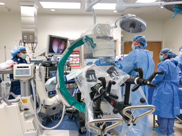
NEW AND IMPROVED The latest C-arms are smaller in size and include several reduced-dosage modes that still capture quality images.  |  Pamela Bevelhymer

The technological enhancements found on newer C-arms are designed to improve radiation safety. Here are some of the features the latest models offer, according to Bart J. Friedman, MD, a radiologist at Allegheny Valley Hospital in Natrona Heights, Pa.:

Laser aimers. This feature improves the accuracy of the visual aiming of the X-ray tube in an exposure-reducing way. Instead of turning on the fluoroscopy and trying to find where you are on a patient, laser aimers help guide you to the area of the body that needs to be imaged before you even turn on the equipment.

Flat-panel image detectors. These produce lower dosages of radiation, and their smaller size allows staff to navigate around the sterile field with more freedom than they could with the older designs.

Last-image hold. With this feature, the last captured image is digitally held or frozen on the monitor after the X-ray has ended. It’s a dose-saving benefit that allows radiologists and surgeons to analyze an image and determine the next step in the procedure without keeping the fluoroscopy activated.

Reduced dosage capabilities. C-arms now come with “low dose” or “pulse” modes that offer providers adequate X-ray images while emitting less radiation.

Jared Bilski

That guiding principle also applies to patient positioning. “Patient exposure to radiation increases exponentially by how close they are to the X-ray tube, so always try to position the patient as far away as possible from the tube,” says Mr. Kekosh. Specifically, he says, the patient should be 12 to 15 inches away from the X-ray tube and positioned as closely as possible to the image intensifier.

Promote PPE use. Even with the proper dosages of radiation in play and patients and staff positioned as far as possible from exposure risks, there are still some risks left to mitigate. That’s where the use of protective equipment comes into play. Staff members who work imaging cases should be properly shielded and keep a close eye for any gaps in protection.

For instance, leaded eyewear should always include side shields. “Without side shields, your eyes are not protected if you look away from the C-arm’s X-ray tube to see the image on the monitor,” says Mr. Kekosh. You should also wear leaded gloves and a wrap-around apron to keep a layer of protection from the X-ray tube, he adds.

In addition to all the gear Mr. Kekosh described, Dr. Friedman touts the importance of radiation badges or dosimeters, which detect and measure the amount of radiation staff has been exposed to over time. At his facility, radiology technologists who frequently work surgical cases are monitored for levels of exposure. “If their quarterly reports exceed safe levels, they’re excused from working cases for a quarter.”

In addition to protective clothing and equipment, structural shielding options help to reduce exposure to radiation scatter, such as lead acrylic shields placed under the OR table or mounted on the ceiling, adds Mr. Kekosh.

In-house experts

Remember, whether it’s training staff on radiation safety and proper image techniques or getting feedback on C-arm purchasing decisions, your best resource will always be your radiologists and radiology technicians. Take full advantage of their expertise on this critical safety issue because, as Dr. Friedman points out, “Other specialties simply don’t have as much training and knowledge on radiation safety.” OSM 

Related Articles

Be an Agile Learner

In a modern workplace where change often comes quickly and relentlessly, a leader should embody the concept of learning to embrace an agile mindset....