The Next Big Thing in Surgical Video

Share:

It's not just larger screens and better resolution, it's also about integrating processes and empowering surgeons and staff.


VIEW FROM THE TOP Ceiling-mounted monitors displaying ultra-high-def video hang around the sterile field in state-of-the-art ORs, leaving the floor clear of cords and wires.   |  John Martin-Eatinger

When most people think of "video," they think screens ?— specifically, their size and whether they have HD or 4K resolution. In an OR, size and resolution are obviously crucial, but to truly leverage the power of surgical video, consider how it can integrate seamlessly into the rest of what you do.

Video integration goes beyond presenting excellent images and video during surgery. It means automatically sending intraoperative images and video to an electronic medical record (EMR). It means enabling providers to communicate, collaborate and visualize what's happening in one OR without leaving another. It means empowering your entire staff by allowing them to see what surgeons see during procedures.

Planning ahead

To implement video integration, focus on your short- and long-term goals. It's a lot of work and involves months of planning, says Jocelyn Chua, RN, BSN, CNOR, executive director of surgical services, GI lab and women's health at Advocate South Suburban Hospital in Hazel Crest, Ill., and Advocate Trinity Hospital in Chicago. As part of a two-year, eight-figure revitalization project completed in 2020, Ms. Chua relished the chance to build nine new ORs with integration capabilities. She says the extensive preparation for the project enabled her team to work well with their eventual video integration vendor. "We were able to give them clear direction as to what we wanted, and they provided us with a video integration program that's able to support our growing robotics program, while also giving us 4K and fluorescence capabilities," she says. Jennifer Milam, MSN, RN, NE-BC, director of perioperative services at Ochsner Medical Complex — The Grove, a recently-opened five-story medical complex and surgical hospital in Baton Rouge, La., also envisioned the possibilities of video integration in its four new ORs when she joined Ochsner. "Surgery has made a massive movement toward minimally invasive procedures, so video technology is absolutely critical to everything we do," she says, noting the research her team put into selecting the right system. "We were very fortunate to get site visits and see what a demo facility looked like, and what top-of-the-line equipment looks like, so we could make an educated decision."

Ms. Milam recommends taking your time and looking at vendors beyond your usual partners, so you don't miss out on more user-friendly options or technology offered only by a certain vendor. Also, get your staff involved in the trialing process. "Make sure the people who do the work get the opportunity to see the tech in action," she says. "As a leader, what I see as bright, shiny and fantastic may not function as well once they put it into practical application."

SURGICAL HIGHLIGHT
Improved Visualization of Viable Tissue
GLOWING REVIEW Surgeons appreciate being able to better identify vital structures during laparoscopic procedures.   |  John Martin-Eatinger

Fluorescence-guided surgery can help surgeons perform safer and more accurate procedures. "Our physicians usually use fluorescence for bowel procedures where they're trying to identify viable tissue," says Jocelyn Chua, RN, BSN, CNOR, executive director of surgical services, GI lab and women's health at Advocate South Suburban Hospital in Hazel Crest, Ill.

Previously the surgeon selected the tissue based on their own experienced eyeballing of the area. Now, the fluorescent agent indocyanine green is injected into the patient and then, before dissection, the surgeon can better identify viable tissue based on the image augmented by the dye on the monitor. Ms. Chua regards this as a safety feature as much as a clinical benefit, as the increased precision reduces risks of postoperative infection and leaking.

— Joe Paone

Space and design

Ms. Milam says video integration wouldn't have been possible in the older, smaller ORs in which she's worked. "Our new ORs are built to accommodate surgical robots and microscopes — things that take up a large footprint," she says. In each OR, two large monitors hang on separate walls, while another two are swinging on boom arms that can be moved around the surgical field for optimal viewing.

The video equipment consumes exactly zero floor space. "And it's all mobile," says Ms. Milam. "If you're performing a procedure that doesn't require video equipment, it can be swung around and moved up against the wall where it's not in the way."

Ms. Chua's old ORs were 400 square feet; the new ones are 625 square feet. Each feature two 32-inch monitors hanging from the ceiling, and a much larger wall-mounted monitor. They're all 4K touchscreens. "The monitors hanging from the ceiling are usually used by the physician and their assistants in the sterile field," she says. "The anesthesiologists and circulators tend to use the one on the wall so they're not hovering behind someone's back in the sterile field to see what's going on."

Infrastructure is also vital to make video integration work. For example, access to power sources and data connections is crucial. Carefully plan where, how many and what types of wall outlets you'll need. Ms. Chua says video signal outlets in her ORs let the surgical team plug and play devices on the fly, a game-changer for the whole staff. The signals can be routed anywhere inside or outside the OR, opening up previously unimaginable conveniences.

"In the past, during procedures that required a microscope, surgeons or their assistants were the only ones who had visibility of the surgical field through the eyepieces," she says. "Now they plug the microscope into a video outlet, and the image can be seen by everybody in the room."

Wires and cords are also accounted for in the design. "You don't see any of the wiring because it's inside a bracket hanging on the wall, and embedded in the boom system," says Ms. Chua. Ms. Milam has a similar setup with ceiling-mounted booms. "All the pieces we need for laparoscopic surgery or any type of video surgery are on the booms," she says. "Floors are free and clear of cords and clutter."

Collaboration and integration

TUNING IN ?Thanks to the new video integration system, Advocate South Suburban Hospital Clinical Pathologist Alia Salhadar, MD, can look live at the same views surgeons are seeing and have a conversation about what looks like viable tissue for dissection.   |  John Martin-Eatinger

Both Ms. Chua and Ms. Milam rave about the ability their video integration systems provide for increased collaboration not only inside the OR, but beyond the walls as well. Videoconferencing in particular is a major benefit. Take, for example, 
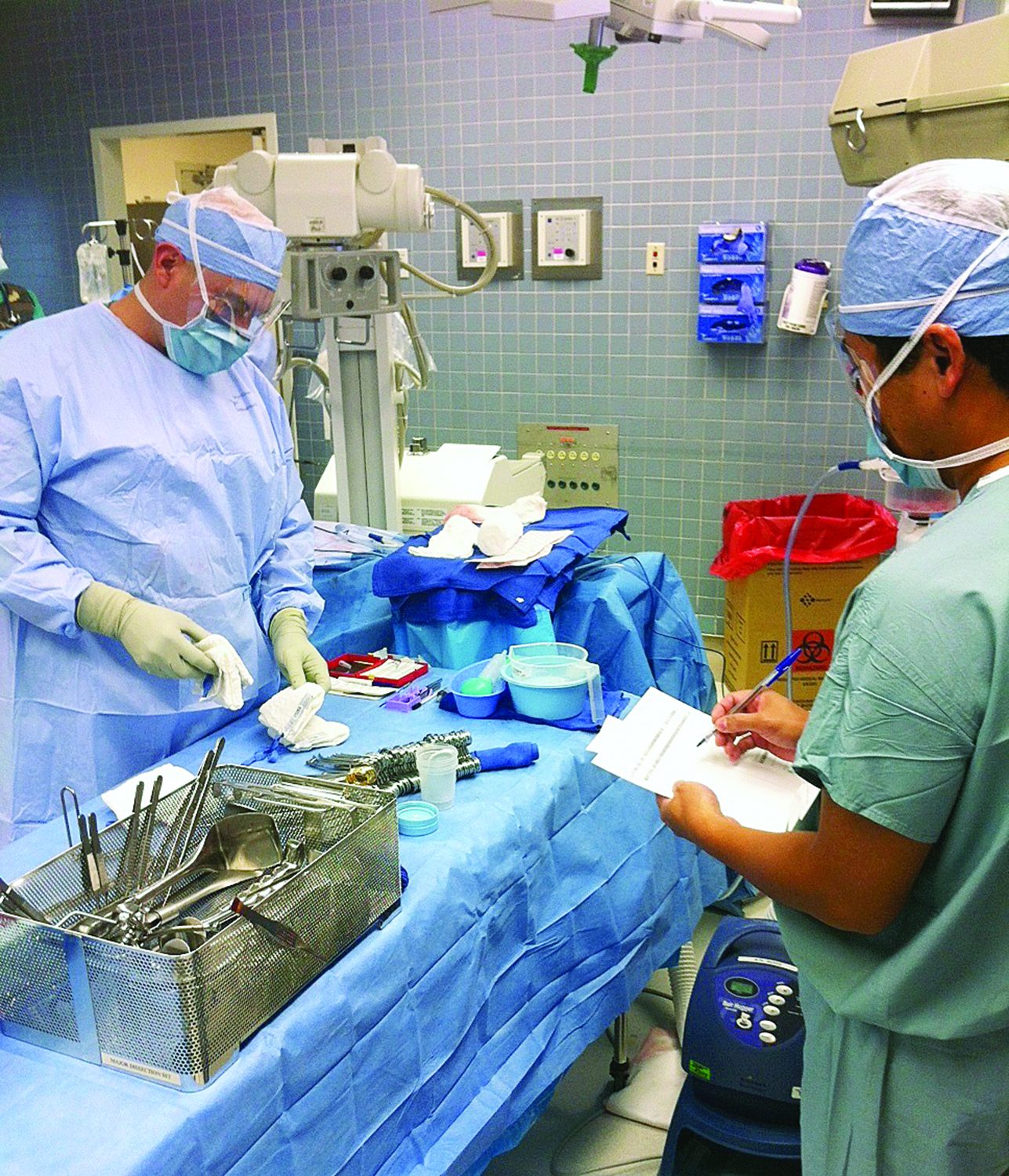
an intraoperative biopsy. Previously, says Ms. Chua, the surgeon would remove tissue and send it to pathology for dissection. Then the pathologist would walk to the OR or use the phone to tell the surgeon if the sample was sufficient or if 
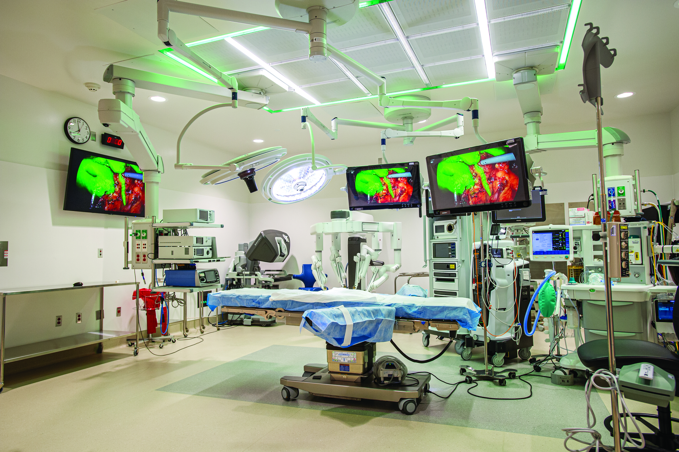
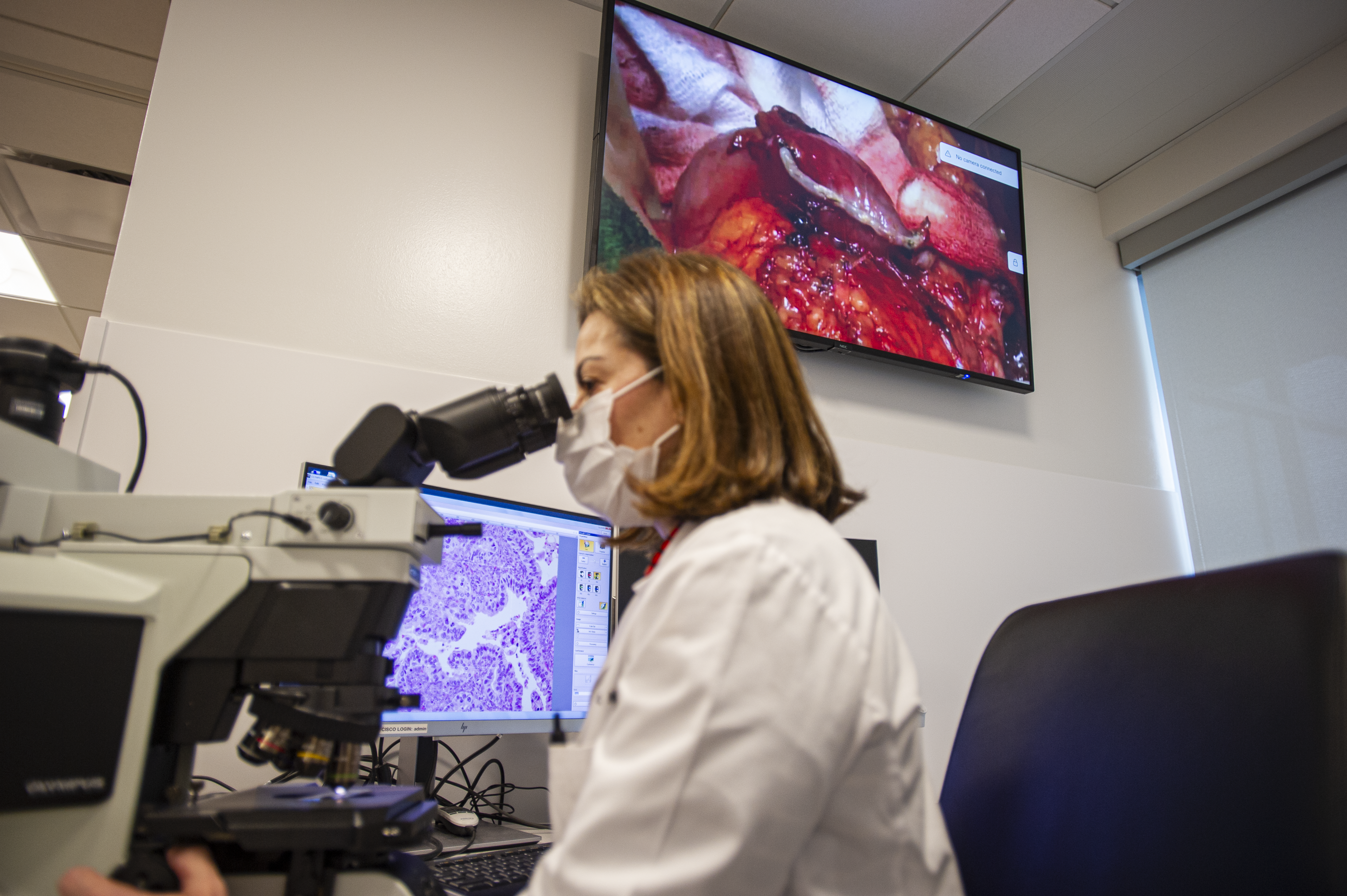
more tissue was needed. Now, thanks to cameras embedded in the OR's surgical lights, the surgeon can simply share image of the surgical field, live, with the pathologist sitting in their lab or office. "The two of them can have a conversation through the speakers in the OR about whether to finish the procedure or continue removing tissue," she says.

At Ms. Milam's facility, surgeons in different ORs can consult while performing surgeries. "For example, a GYN surgeon working in one OR and a general surgeon working in another can share video images in real time on each other's screens and discuss what they see," she says. Her facility has used video integration to show live surgeries in its conference rooms for teaching purposes, too.

In a video integration system, all images and videos are managed and processed through what is essentially a combined server and router. This enables videoconferencing and can deliver those images and videos to EMRs.

This capability has made a big difference at Ms. Chua's facility. Previously, patient info was manually entered to tag images and videos from surgeries, and staff had to print numerous copies of images to add to medical records for the surgeon to evaluate and to share with patients and their caregivers. Now all OR staff need to do is open the patient's EMR.

"From there, it automatically interfaces the video system," says Ms. Chua. "When the surgery is complete and the surgeon is informing the patient's family, they've already pulled all the pictures and annotated them. If they have 20 pictures and only want to give the patient three that reflect the procedure, they just add those images into their discharge notes."

Perioperative professionals must be well-versed in the capabilities and operation of video integration before they use it in live procedures. At Ms. Chua's facility, an in-service was held with a large group, followed by one-on-one trainings for each staff member. She says her tech-savvy staff was off on their own within a week. But it's not just the surgeons and OR staff who need to be comfortable with the new equipment and technology. "We also wanted to make sure our ancillary team and support services were comfortable, because they're the ones who need to do the preparation, upkeep and end-of-day cleaning of the area," she says.

Ultimately, video integration is way more than pretty pictures. It allows surgeons, OR and support staff to work more quickly, intelligently, precisely and collaboratively. OSM

Related Articles