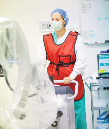
Members of your surgical team are at risk every time C-arms are switched on from a danger they can't see or feel. Luckily, the steps they can take to protect themselves from X-ray scatter are rooted in common sense. My role as a radiation safety officer at a large hospital lets me track the amount of radiation thousands of surgical professionals are exposed to each year, and most have little to extremely low levels of exposure because they work with the latest imaging technologies, practice proper fluoroscopy techniques and understand the 3 fundamentals of radiation safety.
- Home
- Article
Focus on the Fundamentals Of Radiation Safety
By: Michael Bohan
Published: 9/27/2019
Understanding the principles of time, distance and shielding will limit your team's exposure risks.
ALARA ("as low as reasonably achievable") is the guiding principle of radiation practices: use the lowest possible dose to obtain needed images. That can be achieved by keeping the beam-on time to an absolute minimum. The Golden Rule is to reduce fluoroscopy doses to the minimum necessary to achieve the clinical result.
Several features on newer machines help you achieve that aim:
- Pulsed fluoroscopy delivers radiation in short bursts, instead of continuously, to capture images.
- Loop functions cycle through a series of captured images, letting the physician review what has been captured to decide if he can avoid reactivating the C-arm to capture useful images.
- Last-image hold features freeze the image that's captured as soon as the C-arm is deactivated. This mode can be used in place of live fluoroscopy to confirm anatomical information and the locations of hardware and implants.
Remember, doses accumulate as exposure time increases. Relying on these features found on new C-arms and a judicious use of fluoroscopy will limit the amount of time the C-arm is activated.

Radiation intensity drops off rapidly with distance. While physicians and X-ray techs control how long radiation is used in a procedure, the OR staff can control their proximity to it. When a surgeon or tech steps on the pedal of the C-arm, radiation is being produced. There is usually a light on the gantry that goes on when the pedal is pushed. Or you can simply look at the monitor. If you see a moving picture, that means the X-ray machine is on (unless it's in loop review mode). When the light goes on, or the image on the monitor starts to move, take a step — or 2 or 3 steps — back.
Most radiation is absorbed into the patient's body. Sometimes it gets scattered to the side, though, and this scatter is what you need to be concerned about. There are several ways to limit radiation scatter:
- Collimation. Collimating the X-ray beam to focus on the intended imaging site reduces the dose delivered to the patient and improves image quality due to a reduction in X-ray scatter.
- Proper positioning. Place the X-ray tube underneath the OR table, as far from the patient as possible, and position the image intensifier as close to the intended imaging site. Standing near the image receptor side of the C-arm, where radiation scatter is less than near the X-ray tube, will limit your level of exposure.
If the C-arm is positioned vertical, or near vertical, keep the X-ray tube under the patient. (The exception to this is when mini C-arms are used during extremity procedures. The dose rate is low enough that it's more practical to have the X-ray tube above the surgical field.)
- Flat-panel detectors found on newer C-arms capture higher quality images at lower radiation doses, which limits scatter and exposure risks to staff and physicians.
The intensity of scatter radiation is much less than that of the beam that enters the patient. For example, the dose rate going into a patient undergoing an abdominal fluoroscopy is about 20 millisieverts per minute, but the dose rate 1 foot away from where the beam is traversing the body laterally ?— about the distance where somebody would be standing next to surgical table — is about .3 millisieverts per hour. That's a reduction by a factor of 60 from the patient's dose. Using the Inverse Square Law, if your exposure is .3 millisieverts at 1 foot, you can reduce that exposure by 75% simply by standing 2 feet away and by 89% if you stand 3 feet away.
Every member of the surgical team should wear lead aprons, which protect wearers against 95% of radiation scatter. Staff members who need to be adjacent to the OR table should also wear thyroid shields and leaded glasses. Eye and thyroid protection are optional for staffers in the room who generally are more than 6 feet away from the table.
Got a question about imaging safety? Radiation professionals can be consulted at the Health Physics Society at hps.org. Click on the "Ask the Experts" section at the top of the website, where you can browse answers from previously asked questions or submit a new one. Within a week, an expert from your region will post an answer.
The annual allowable radiation exposure is 50 millisieverts. The ALARA Level 1 threshold is 5 millisieverts — 10% of the annual limit. That's the pay-attention threshold. The ALARA Level 2 threshold is 15 millisieverts, or 30% of the annual limit. That's the investigational threshold where you're going to have to find out what your team has been doing to get that kind of exposure, which shouldn't be happening if they've been following good safety practices.
Here at Yale New Haven Hospital, which has thousands of operating room staff members, we give radiation dosimeters — which measure exposure levels — to only about 1,000 workers, the ones who we think are most likely to have radiation exposure of any significance. Smaller hospitals and freestanding ambulatory surgery centers don't have professional health physics experts on staff, so they often give dosimeters (also called film badges) to all members of the clinical team.
Dosimeters are retrospective tools that allow you to assess the effectiveness of your radiation safety program. If you're following good practice, the vast majority of your employees won't be getting exposed to high amounts of radiation, so the results from the dosimeters should be negligible. In lieu of having professionals on hand to determine who needs these devices, it's easier to badge everyone who works imaging cases.
Maintain perspective
We're all exposed to about 3 millisieverts of natural radiation each year. The effective dose of a chest CT is about 5 millisieverts, which is equivalent to about a year and change of our natural exposure. Yes, you're working with radiation, but if you understand the fundamentals of time, distance and shielding, you can easily keep your occupational exposure well within the variation we're exposed to by simply walking outside. OSM
.svg?sfvrsn=be606e78_3)
.svg?sfvrsn=56b2f850_5)