Your Road to Safer Lap Choles

Share:

Strategies to prevent bile duct injuries during gallbladder removal.


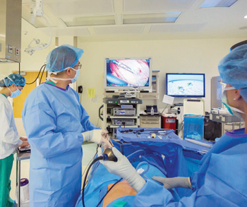
For all the benefits of laparoscopic cholecystectomy, a persistent problem continues to plague one of the most commonly performed surgical procedures: bile duct injuries (BDIs). This dreaded complication can turn an otherwise routine same-day gallbladder removal into an emergency — and a medical malpractice suit. BDIs are the No. 1 litigated case against surgeons, responsible for 1 in 5 malpractice claims.

What is the frequency of BDIs? It's relatively rare — it's estimated that only 0.4% of lap chole complications results in BDIs — but that translates to about 3,000 nicked, burned or pinched bile ducts per year when you consider that surgeons laparoscopically remove 750,000 to 1 million gallbladders annually. BDIs lead to symptoms that can be painful, even deadly, if not treated. Here are tips to make a safe operation safer.

One approach toward improving safety is standardizing dissection of the gallbladder during surgery by creating a "critical view of safety" — a technique endorsed by the Society of American Gastrointestinal and Endoscopic Surgeons (SAGES). SAGES suggests these 6 strategies that surgeons can employ to adopt a universal culture of safety for cholecystectomies that minimizes the risk of BDIs.

1. Critical View of Safety.

Use the Critical View of Safety (CVS) method to identify the cystic duct and cystic artery during lap choles. The 3 criteria required to achieve the CVS are:

  • clear the hepatocystic triangle — the triangle formed by the cystic duct, the common hepatic duct and the inferior edge of the liver — of fat and fibrous tissue;
  • separate the lower one-third of the gallbladder from the liver to expose the cystic plate, also known as the liver bed of the gallbladder; and
  • see 2 and only 2 structures when entering the gallbladder.
BETTER VISUALIZATION
Overcoming 'Visual Perceptual Illusions'
CRITICAL VIEW Visual misperception causes most of the errors that result in laparoscopic bile duct injuries, not errors of skill, knowledge or judgment.

The primary cause of error in the vast majority of BDIs? Not poor technical skills or a lack of knowledge on the surgeon's part, but visual perceptual illusions, meaning the surgeon deliberately cut the common duct, erroneously believed to be the cystic duct. Here are a couple ways to improve visualization.

  • ICG exposure. An injection of indocyanine green (ICG) dye just before you start the case can help. Once you inject ICG, near infrared imaging picks it up as it illuminates structures such as the cystic duct and the common bile duct. In some cases, so-called "ICG exposure" lets you better visualize the biliary tract so you can safely transect the cystic duct and avoid any injury to the common bile duct.
  • Operative cholangiogram. In an intraoperative cholangiogram, you transect the cystic duct, see a catheter through the cystic duct into the common bile duct and then inject a radio opaque contrast material. Cholangiograms let you visualize the common bile duct stones. There are drawbacks to this technique: it takes more time, a little more equipment, and it exposes you and your team to radiation.

- Salvatore Docimo Jr., DO, FACS

2. Aberrant anatomy.

Understand the potential for aberrant anatomy in all cases. Aberrant anatomy may include a short cystic duct, aberrant hepatic ducts or a right hepatic artery that crosses anterior to the bile duct.

3. Imaging.

Make liberal use of cholangiography or other methods to image the biliary tree intraoperatively. Cholangiography may be especially important in difficult cases or unclear anatomy. Studies have shown that cholangiography reduces the incidence and extent of bile duct injury, but the subject remains controversial.

4. Time out.

Take an intraoperative time out during a lap chole before clipping, cutting or transecting any ductal structures. The time out should consist of a stop point in the operation to confirm that you've achieved the CVS.

5. Dangerous dissections.

Recognize when the dissection is approaching a zone of significant risk and halt the dissection before entering the zone. Finish the operation by a safe method other than cholecystectomy if conditions around the gallbladder are too dangerous. In situations where there is severe inflammation in the porta hepatis and neck of the gallbladder, the CVS can be difficult to achieve. The fact that achieving the CVS appears not feasible is a key benefit of the method, as it alerts you to possible danger of injury.

6. Ask for help.

Get help from another surgeon when the dissections or conditions are difficult. When it is practical to obtain, the advice of a second surgeon is helpful under conditions in which the dissection is stalled, the anatomy is unclear or under other conditions deemed difficult by the surgeon.

Continually improving techniques

Though rare, the reality is that BDIs still occur during lap choles. Any technique or technology that can help eliminate the risk of injury to the common bile duct structure is worth considering. OSM

Related Articles