Thinking of Buying C-arms

Share:

A look at how the newest platforms capture and store high-quality images.


C-ARM STRONG Advanced technology has made C-arms safer and more efficient so that your surgeons can provide their patients with the best possible care.

It's hard to believe that I used to perform spine surgery by moving instruments based on feel, my ability to recall specifics about the patient's anatomy and grainy pictures captured by big, bulky C-arms. Those early days of intraoperative imaging didn't help lower my stress level while I worked around such delicate anatomy, where the difference between excellent outcomes and devastating complications is measured in millimeters. Thankfully, newer C-arms have taken much of the guesswork — and some of the mental strain — out of the challenging cases. Starting below, we highlight 8 of the newest models.

Orthopedic and spine surgeons can now magnify and enhance high-definition images without losing an ounce of clarity, meaning they can administer spinal injections for pain management more precisely and double-check the placement of hardware and implants long before patients leave the OR. Newer C-arms also require lower radiation doses to capture higher quality images, a feature that helps protect staff and surgeons from imaging's invisible danger.

The latest C-arms can take big chunks out of your capital equipment budget — basic models generally cost $100,000 to $125,000, while platforms with bells and whistles run between $300,000 and $350,000 — so consider these factors to make smart purchasing decisions while investing in units that will provide your surgeons with the enhanced intraoperative images they want and need.

  • Image quality. The latest C-arms boast flat screen monitors, which display clear, crisp ultra-high definition images of targeted anatomy. Many manufacturers are swapping out conventional image intensifiers for flat-panel, digital image detectors, which provide higher-quality images and a wider field of view with less distortion at a fraction of the radiation output.

Those features match imaging's fundamental principle: capture quality images with radiation doses that are as low as reasonably achievable (ALARA). In other words, use the lowest possible dose to capture images with enough detail to guide the surgeon's work. Radiation scatter still occurs with digital image detectors, but because a lower dose is used to begin with, the scatter effect is minimized, and exposure risks are therefore far less for everybody in the room.

  • Data storage and retrieval. Today's C-arms have the capacity to store many years' worth of images, but most facilities invest in a more limited (less expensive) storage capacity and download images daily, weekly or monthly to a picture archiving system. Surgeons can also download images to an encrypted flash drive for later viewing.

C-ARM SAFETY Digital image detectors minimize radiation scatter, protecting your staff and patients from an invisible — and cumulative — danger.

That's a nice feature to have, and one I've used to improve patient care. For example, when a patient with chronic back pain tells me the first in a series of medication injections effectively eased his discomfort, I call up the digital images captured during the procedure to review exactly where I placed the injection. On the other hand, if the injection was ineffective, I reassess the site and technique to determine how I can improve the patient's treatment moving forward.

  • Space-saving design. Digital image detectors are less bulky than conventional image intensifiers, making the newest C-arms more compact than previous generations. They take up less room in crowded ORs and are easier to maneuver in the tight confines of the sterile field. That increased maneuverability helps you position the image detectors at optimal angles around the patient to capture high-quality images of targeted anatomy.

C-arms are complex machines with a lot of moving parts, so be sure the manufacturer of the unit you purchase includes a comprehensive annual service contract. And know what you're buying before making an investment. Most vendors will provide you with the C-arm you're considering for a one-week trial, while some will extend the trial period to a month. It's important to get feedback from as many surgeons as possible during the trial period, because they're the ones who ultimately want access to high-quality intraoperative images so they can perform safer and more effective procedures with added confidence. OSM

Carestrea\m

Carestream

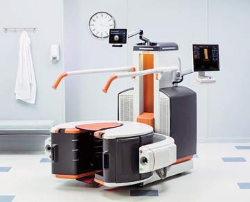
OnSight 3D Extremity System

carestream.com
888-777-2072

Price: not disclosed

FYI: This standalone, compact CT unit fits in a standard clinic exam room and is designed to help orthopedic surgeons diagnose the location and severity of upper and lower extremity injuries. The tilt, height and rotation of the unit's flat-panel image detector are easily adjustable, and intuitive touchscreen controls guide staff through the process of capturing 3D images of weight-bearing and non-weight-bearing joints. The high-quality scans let surgeons assess fractures and joint injuries in greater detail in order to make more informed clinical decisions. The unit can be readied for use in 5 minutes and scans are completed in 10 minutes, says the company.

CurveBe\am

CurveBeam

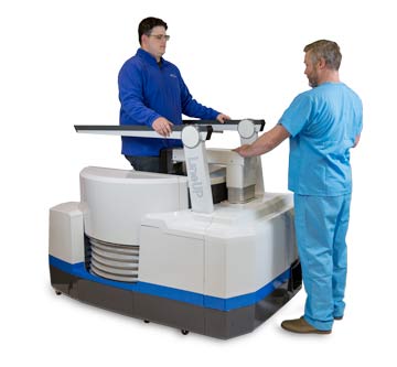
LineUp System

curvebeam.com
267-483-8081

Price: $225,000

FYI: Use this standing CT unit to capture bilateral 3D scans of weight-bearing feet, ankles and knees for the most accurate assessments of bone alignment and joint spaces. Scan time is less than 30 seconds and the required radiation dose is virtually equivalent to a standard X-ray exam, says the company. Automatic metal artifact reduction and a large field of view provide realistic high-resolution 3D images of joints, even those that are filled with implanted hardware from previous procedures.

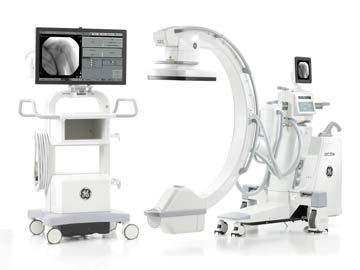
OEC Elite \CFD

GE Healthcare

OEC Elite CFD

gehealthcare.com
866-281-7545

Price: not disclosed

FYI: The OEC Elite CFD features a flat-panel image detector designed to capture quality images at a lower radiation dose. Its continuous fluoroscopy capability provides detailed views of anatomy without the distraction of stuttering, visual lag or ghosting. The unit provides a large field of view that lets you see 20% percent more anatomy during procedures, says the company. Staff can use a handheld remote or table-side controls to easily control the unit's movement and position the flat-panel image sensor at the angles needed to obtain anatomical views quickly and easily.

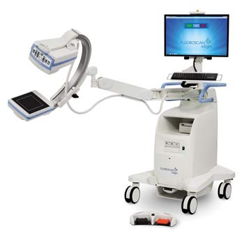
Fluoroscan Insight FD Mini C\-arm

Hologic

Fluoroscan Insight FD Mini C-arm

hologic.com
508-263-2471

Price: $70,000 to $90,000

FYI: The range of motion of this mini C-arm lets staff position the unit's rotating flat-panel image detector at angles that capture distortion-free images of the patient's extremities. A low-dose mode reduces radiation output by up to 34%, says the company. Buttons at the tube head let the surgical team control the arm from the sterile field, and intuitive touchscreen controls on the unit's 24-inch monitor let support staff adjust the dose rate and resolution of captured images. The monitor can be tilted and swiveled into the best viewing angle for surgeons and staff.

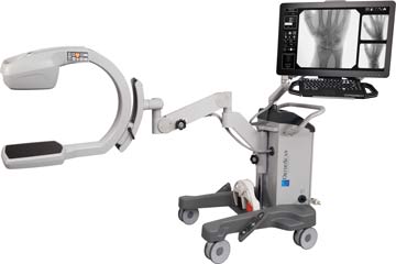
OrthoScan FD \Pulse

OrthoScan

OrthoScan FD Pulse

orthoscan.com
480-503-8010

Price: not disclosed

FYI: The pulsed fluoroscopy feature on this mini C-arm reduces the radiation dose needed to capture high-quality images. Its 15 cm x 15 cm flat-panel image detector, the largest available on any mini C-arm, provides a wide field of view, says OrthoScan. The unit's 24-inch high-resolution monitor displays detailed images of target anatomy, and staff can scroll through the monitor's intuitive touchscreen controls much like they would on their personal smartphones or tablets. Surgical LED lights on the tube head are designed to reduce shadows on the surgical field and improve the surgeon's view of surgery.

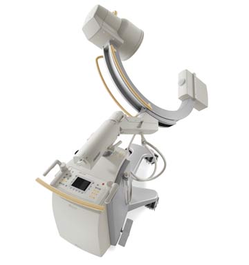
BV P\ulsera

Philips

BV Pulsera

usa.philips.com
800-229-6417

Price: not disclosed

FYI: The system's 9-inch or 12-inch image intensifier captures high-quality images during complex orthopedic cases, including minimally invasive spine and hip procedures. Its Ortho Plus mode boosts the imaging power when it's needed to penetrate dense anatomy on obese patients or during challenging scenarios, such as imaging of the lateral hip. Integrated BodySmart software defines the optimal imaging field and automatically adjusts imaging parameters to provide the best possible image quality, says Philips. Height-adjustable and rotatable dual display monitors are housed on a mobile viewing station for placement in optimal viewing locations around the surgical table.

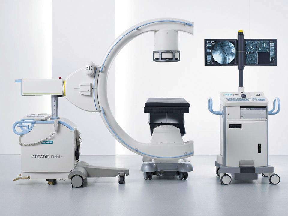
Arcadis O\rbic 3D

Siemens USA

Arcadis Orbic 3D

healthcare.siemens.com
888-826-9702

Price: not disclosed

FYI: The arm of this unit rotates 190 degrees around the patient to provide precise 3D images of targeted anatomy, says Siemens. Its isocentric design — ?the distance between the image intensifier and target anatomy remains constant as the C-arm rotates — maintains consistency in the size of captured images, even as the arm rotates through various angles. Proprietary image processing technology analyzes captured images and automatically adjusts the dose, brightness and contrast to optimize the image's clarity. Task menus let you toggle through preprogrammed settings, such as procedure type and body part to be scanned, with the click of a mouse.

Ziehm Imaging

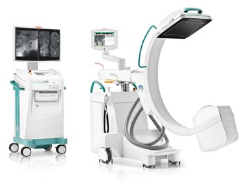
Ziehm Vision RFD 3D

ziehm.com/RFD3D
800-503-4952
+49 911 21 72 0

Price: not disclosed

FYI: This system's SmartScan software combines linear and rotating movements of the C-arm to capture 180-degree 3D scans of targeted anatomy at every point along the field of view. Its SmartDose software, which automatically adjusts pulse rate and width based on the anatomical area to be imaged, is designed to limit dose exposure for patients and staff, says Ziehm. Easy-to-understand icons on a touchscreen monitor let staff control movements of the C-arm and manage what's displayed on the platform's flat-screen monitor. Staff can use the system's integrated workflow wizards to set up optimal settings for the scanning of specific body parts in about 2 minutes. The unit also boasts a small footprint, wireless video capabilities that send images to external monitors and bedside controls that let staff prep the unit for use from the sterile field.

Related Articles