CRNAs Focus on Staff Wellness and Patient Safety
The American Association of Nurse Anesthesiology (AANA) has joined the ALL IN: Wellbeing First for Healthcare coalition, saying the group’s initiative to improve the...
This website uses cookies. to enhance your browsing experience, serve personalized ads or content, and analyze our traffic. By clicking “Accept & Close”, you consent to our use of cookies. Read our Privacy Policy to learn more.
By: Daniel Cook
Published: 4/15/2019
The next time you need a lesson in radiation safety, pop your head into a procedure room to watch an interventional pain doc at work.
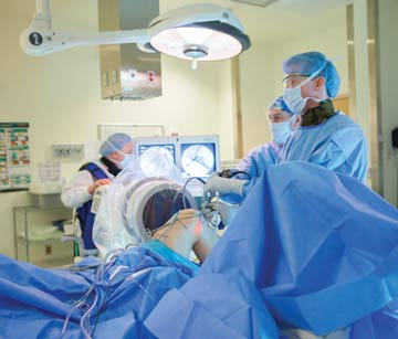
“They’re in front of C-arms all day long, so they respect imaging’s invisible dangers,” says Daniel Kaplan, DO, an orthopedic surgeon at WellSpan Orthopedics, York, Pa. “They step back from the imaging field and clear their hands during every shot, and they always wear the appropriate personal protection equipment. You can tell they’ve been fully educated on radiation safety and receive continuous training.” Your surgeons and staff know the basics of radiation safety, but they might not fully appreciate the risks they face every time a C-arm is wheeled into the OR. No one on your team would consider working a case involving intraoperative imaging without wearing a lead apron, but do they always don leaded eye protection and thyroid shields? They should.
“The thyroid and eyes are extremely sensitive to chronic radiation exposure, which is believed to cause most papillary thyroid cancers and can lead to the formation of cataracts,” says Dr. Kaplan. “Surgeons understand that the eyes are vulnerable to radiation, but current options are bulky and uncomfortable, and they tend to fog up. I don’t wear mine consistently.”
If Dr. Kaplan, who has conducted extensive research on intraoperative radiation safety, isn’t fully compliant with wearing proper personal protection during procedures, where does that leave members of your surgical team? Perhaps a perceived lack of practical comfort, not awareness, might be causing them to go without the protection they need. That’s no longer a valid excuse, however.
“The latest versions of protective equipment are much improved, and much easier to wear,” says Michael Groover, DO, a reconstructive orthopedic surgeon at the Cleveland (Ohio) Clinic Foundation. “There’s no reason it shouldn’t be readily available — and used consistently.”
He says new leaded eyewear is designed for a better ergonomic fit, and the latest lead-equivalent full-body protective gear is lightweight and more comfortable than the heavy, standard-issue lead aprons. Also consider providing surgeons and staff with custom-fitted vest-skirt combinations that distribute the weight of the garments and provide full protection without putting added strain on backs and joints.
“That makes a huge difference during long cases,” says Dr. Kaplan. “I’m also much more likely to take better care of my customized garments, to store them properly after use, than aprons taken from the general stock.”
Dr. Kaplan and his colleagues in the OR wear a dosimeter badge on the thyroid shield and another on the inside of the protective vest, against the chest, to measure their levels of radiation exposure. The maximum annual dose surgical team members should be exposed to is 20 mSv for the body. The annual threshold is higher for the thyroid and eyes (150 mSv), and hands (500 mSv). Each month, monitor and document individual dosimeter readings to ensure team members remain under the annual threshold of acceptable exposure to radiation.
The ALARA principle — maintaining radiation doses “as low as reasonably achievable” when capturing intraoperative images — should guide C-arm use in the OR. So should the inverse square law, which states the magnitude of exposure to radiation scatter is inversely proportionate to the distance from the source. Per the law, doubling the distance from the X-ray tube reduces your exposure to scatter by one-fourth. Mindful that most radiation scatter occurs between the X-ray tube and the patient, Dr. Kaplan offers this advice:
To further limit exposure, use low-dose fluoroscopy — pulsed instead of continuous shots — to capture images and activate the C-arm’s collimator to narrow the radiation beam, says Dr. Kaplan. He also points out that high-definition screens on newer C-arms provide clearer images of anatomy and hardware, therefore decreasing the number of pictures surgeons need to take. Clear communication between the surgeon and the tech who’ll position the C-arm throughout a case is essential, says Dr. Kaplan. A 30-second conversation before the case will ensure they’re on the same page regarding the images they need to capture and the order in which they need to capture them.
Dr. Kaplan recommends blocking out the C-arm positions needed to capture multiple images — much like movie directors do with actors to find the perfect camera angles — and placing tape marks on the floor to guide the tech as she adjusts the C-arm during the case. That pre-op planning increases surgical efficiencies and also limits the amount of radiation the surgical team is exposed to during a case.
For example, Dr. Kaplan says it’s challenging to capture lateral images of the hip that clearly show the femoral neck. “The amount of fluoroscopy that’s used will be significantly reduced if surgeons and techs plan ahead and communicate about placing the C-arm in ways that ensures those images are captured without using multiple shots,” he says.
Kern Singh, MD, a professor of orthopedic surgery at Rush University Medical Center in Chicago, touches on the double-edged sword of intraoperative imaging. “It’s risky, but it drives decision-making in the OR,” he says. “The key is to minimize exposure risks.”
Dr. Singh believes developing technology will eventually help meet that goal. “Investments in advanced imaging, including navigation-based technologies and robotics, are being made to limit fluoroscopy use in the OR,” he says. “New flat-panel C-arms also provide 3D images that surgeons can use to confirm the placement of screws and implants, which can play a key role in radiation-reduction strategies.
“The technology is good,” he adds, “but needs to get better before it’s used routinely.”
Until it does, focus on the basics of radiation safety.
“Surgical professionals have a general understanding that radiation safety is an important issue, but there needs to be more of an emphasis placed on ensuring they know where exposure risks are greatest,” says Dr. Groover. “They also need to fully grasp specific measures that can limit fluoroscopy exposure and protect body parts that are most sensitive to radiation.” OSM
The American Association of Nurse Anesthesiology (AANA) has joined the ALL IN: Wellbeing First for Healthcare coalition, saying the group’s initiative to improve the...
Once you understand that being a perfectionist can be harmful to your personal outlook, health and anxiety level, you can unravel what to do about it....
The start of the year is a great time to clear the decks mentally and review for yourself what works and what does not....